IGF-1 LR3 is a modified analog of insulin-like growth factor 1 with a dramatically extended half-life and enhanced potency. The LR3 modification — an arginine-to-glutamic acid substitution at position 3 plus a 13-amino acid N-terminal extension — reduces binding to IGF binding proteins (IGFBPs) by over 100-fold. The result: a half-life of 20-30 hours instead of minutes, and roughly 2-3x greater biological activity than native IGF-1.
This article ranks five IGF-1 LR3 benefits by evidence quality. An important caveat upfront: most research covers IGF-1 broadly, not the LR3 analog specifically. Where evidence comes from animal models or in vitro work, that distinction is noted clearly. The LR3 modification amplifies IGF-1's effects, but direct human clinical data for this specific analog remains limited.
How IGF-1 LR3 Works
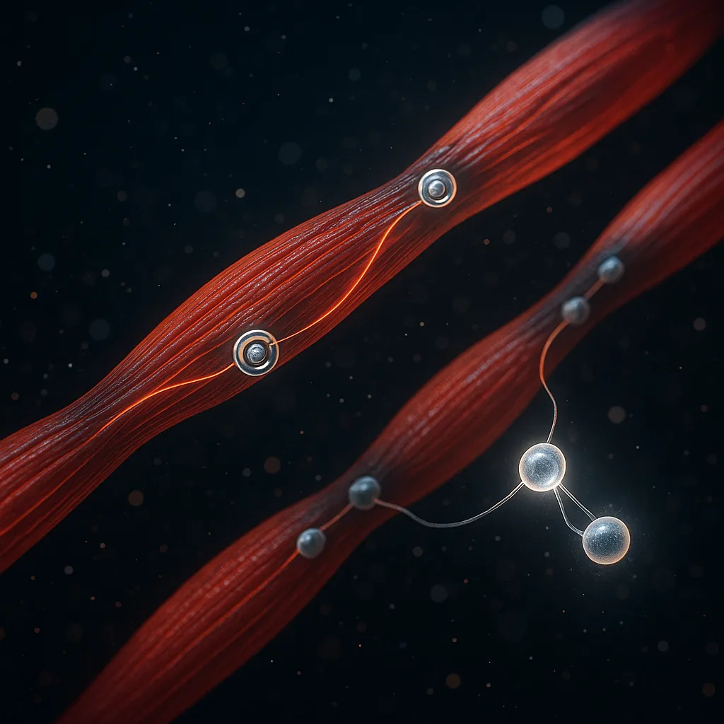
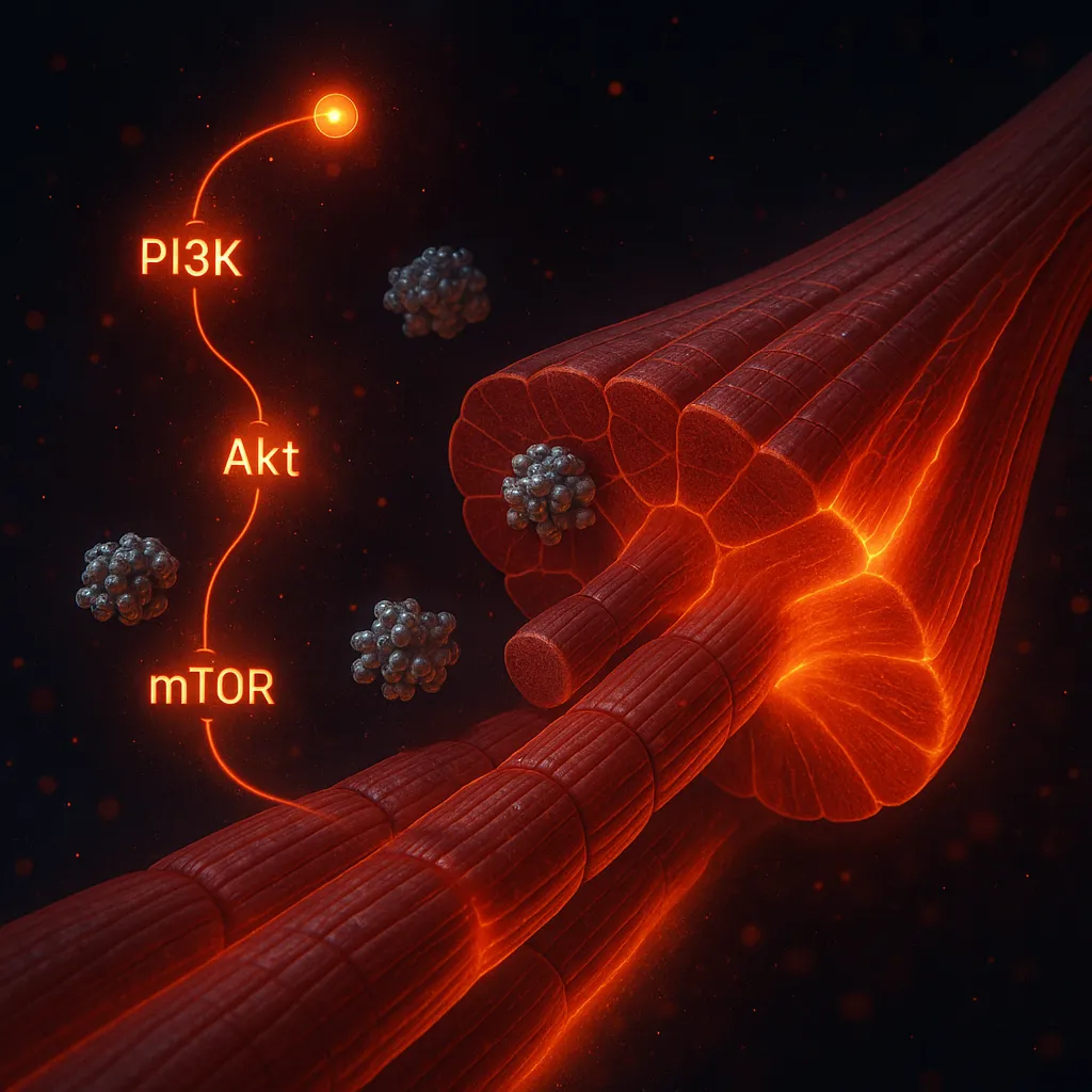
Native IGF-1 is produced primarily by the liver in response to growth hormone stimulation. It circulates bound to IGF binding proteins (primarily IGFBP-3), which regulate its availability and half-life. When IGF-1 binds to the IGF-1 receptor (IGF-1R) on target tissues, it activates the PI3K/Akt/mTOR signaling cascade — the primary driver of protein synthesis in skeletal muscle (Yoshida & Bhatt, 2019).
IGF-1 LR3 bypasses this binding protein regulation. Because it binds poorly to IGFBPs, it remains free and biologically active in circulation far longer than native IGF-1. In animal models, LR3 IGF-1 was approximately 2.5-fold more potent than native IGF-1 in promoting growth, with just 44 mcg/day of LR3 producing effects equivalent to 278 mcg/day of IGF-1 (Tomas et al., 1993).
The downstream effects are broad: increased protein synthesis, satellite cell activation, suppressed protein degradation, nutrient partitioning toward muscle, and tissue repair signaling. For detailed protocols, see the IGF-1 LR3 dosing guide.
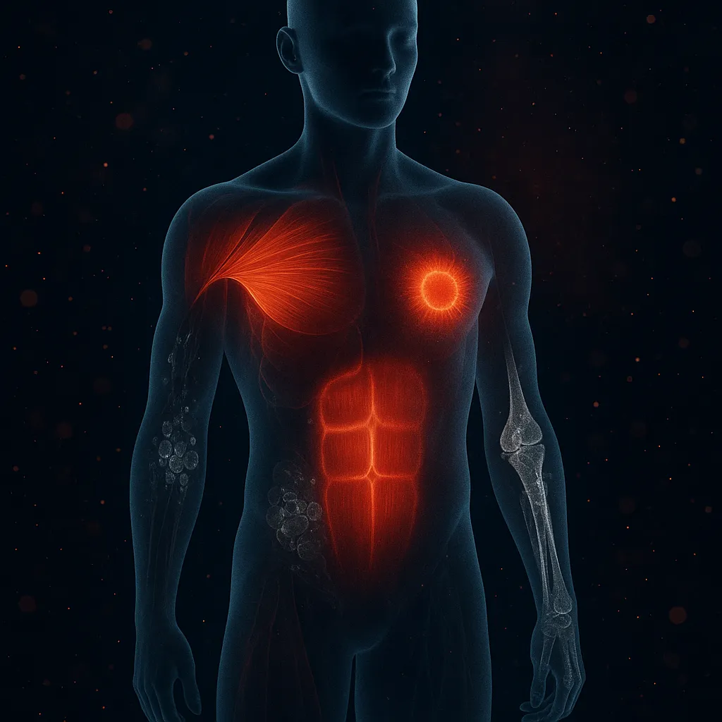
1. Muscle Hypertrophy (Strong Evidence)
The strongest evidence for IGF-1 LR3 centers on skeletal muscle growth. IGF-1 drives hypertrophy through two complementary mechanisms: increased protein synthesis via the PI3K/Akt/mTOR pathway and satellite cell activation that adds new myonuclei to growing muscle fibers.
Adams and McCue demonstrated that localized IGF-1 overexpression in mouse hindlimb muscles produced significant increases in muscle mass, with satellite cell contribution essential to the hypertrophic response (Adams & McCue, 1998). A comprehensive 2020 review confirmed that IGF-1 activates protein synthesis through PI3K/Akt/mTOR and PI3K/Akt/GSK3B pathways while simultaneously inhibiting protein degradation via FoxO suppression (Yoshida & Delafontaine, 2020).
The LR3 modification makes this more potent. In normal growing rats, LR3 IGF-1 produced increased body weight gain, improved nitrogen retention, and greater food conversion efficiency at doses roughly one-sixth those required for native IGF-1 (Tomas et al., 1993).
Evidence quality: Strong for IGF-1 mechanism; moderate for LR3-specific potency (animal data). No controlled human hypertrophy trials with LR3 IGF-1.
Practical takeaway: Muscle growth is the most well-supported benefit. The standard community protocol is 50 mcg/day for 10 days, typically injected post-workout.
2. Anti-Catabolic Protection (Strong Evidence)
IGF-1 does not just build muscle — it actively prevents muscle breakdown. This dual action (anabolic + anti-catabolic) distinguishes it from compounds that only stimulate protein synthesis.
Sacheck et al. showed that IGF-1 suppresses protein degradation by rapidly reducing expression of atrogin-1 (within 1 hour) and MuRF1, two ubiquitin ligases that drive muscle protein breakdown. This occurs through the PI3K/Akt pathway, which blocks FoxO-mediated transcription of these atrophy genes (Sacheck et al., 2004).
This anti-catabolic effect is particularly relevant during caloric deficits, recovery from injury, or periods of forced inactivity — situations where muscle loss accelerates. IGF-1 LR3's extended half-life means this protective signaling persists for 20-30 hours per dose rather than minutes.
Evidence quality: Strong. Multiple studies confirm the mechanism in both cell culture and animal models. The pathway is well-characterized.
Practical takeaway: IGF-1 LR3 may help preserve muscle during cutting phases or recovery periods. This is why some users time cycles around periods of higher catabolic stress.
3. Recovery and Wound Healing (Moderate Evidence)
IGF-1 plays a documented role in tissue repair. A 2021 systematic review of 11 studies found that IGF-1 consistently promoted wound healing by stimulating keratinocyte migration, enabling wound epithelialization, and promoting wound bed contraction. However, only 2 of the 11 studies were conducted in human subjects (Balaji et al., 2021).
IGF-1 also potentiates skeletal muscle regeneration through satellite cell activation — the same mechanism that drives hypertrophy also accelerates repair of damaged muscle fibers (Yoshida & Delafontaine, 2020).
The LR3 modification's extended activity window theoretically provides sustained repair signaling. However, most wound healing research uses native IGF-1, not LR3 specifically.
Evidence quality: Moderate. Animal and in vitro evidence is consistent, but human data is limited. No wound healing trials with LR3 IGF-1 specifically.
Practical takeaway: Recovery enhancement is a secondary benefit. Users stacking IGF-1 LR3 with healing peptides like BPC-157 report faster recovery, though this is anecdotal.
4. Fat Metabolism and Body Composition (Moderate Evidence)
IGF-1 LR3 is not primarily a fat loss compound, but it influences body composition through several mechanisms. The GH/IGF-1 axis has well-documented effects on adipose tissue metabolism, with clinical trials showing reductions in visceral adipose tissue when IGF-1 signaling is enhanced (Berryman et al., 2013).
IGF-1 acts as an insulin sensitizer and improves nutrient partitioning — directing calories toward muscle rather than fat storage. In animal models, LR3 IGF-1 improved food conversion efficiency, meaning more of the consumed calories were directed toward lean tissue growth rather than fat deposition (Tomas et al., 1993).
However, IGF-1 does not directly stimulate lipolysis the way growth hormone does. Its effects on fat are indirect: better nutrient partitioning, improved insulin sensitivity, and increased energy expenditure from greater muscle mass.
Evidence quality: Moderate for body composition changes through the GH/IGF-1 axis. Weak for direct fat loss from LR3 specifically.
Practical takeaway: Do not use IGF-1 LR3 primarily for fat loss. The body composition benefits are a secondary effect of improved nutrient partitioning and increased lean mass.
5. Organ and Tissue Growth (Moderate Evidence — Caution Required)
LR3 IGF-1 infusion in guinea pigs significantly increased the weight of adrenals, gut, kidneys, and spleen while suppressing endogenous IGF-1 and IGFBP concentrations (Bastian et al., 1993). This demonstrates that LR3 IGF-1 drives growth across multiple tissue types, not just skeletal muscle.
This is both a potential benefit and a serious risk. For individuals recovering from gut damage or tissue injury, the broad anabolic signaling could theoretically support repair. But uncontrolled organ growth — particularly gut distension (sometimes called "GH gut" in bodybuilding) and potential cardiac hypertrophy — is a well-documented concern with prolonged growth factor elevation.
Evidence quality: Moderate. Animal data clearly demonstrates the effect. The concern is that this "benefit" becomes a liability with excessive dosing or prolonged cycles.
Practical takeaway: This is why cycle length matters. The standard 10-day on / 4-week off protocol limits exposure and reduces the risk of unwanted organ growth. Never run extended cycles without medical supervision.