Best Peptides for Inflammation: 5 Ranked (2026)
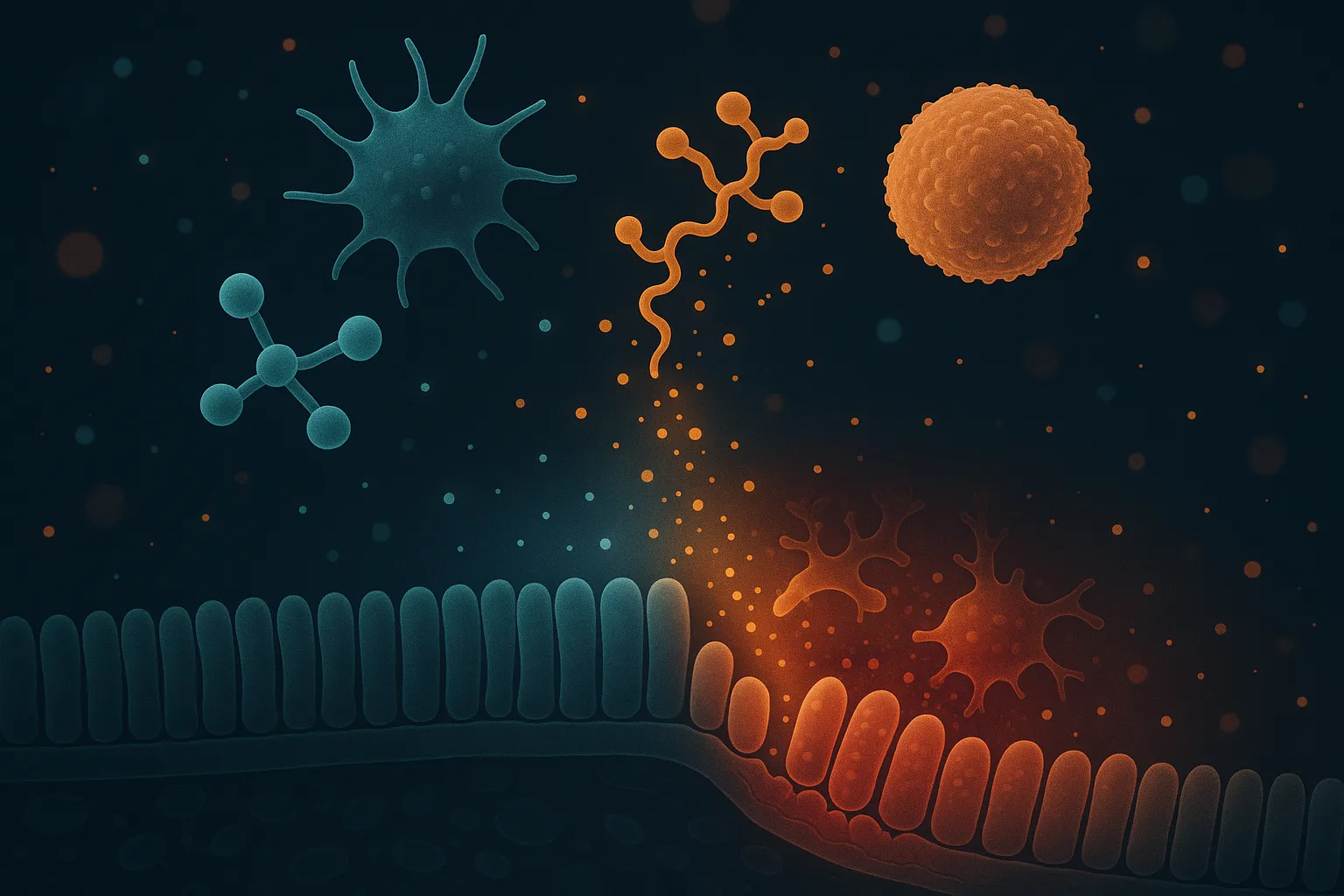
Chronic inflammation is the upstream driver of most degenerative disease: joint destruction, cardiovascular damage, neurodegeneration, metabolic dysfunction, and accelerated aging all share sustained inflammatory signaling as a common mechanism. Conventional anti-inflammatory drugs -- NSAIDs and corticosteroids -- provide symptom relief but carry significant long-term risks and do not address the underlying immune dysregulation.
Anti-inflammatory peptides work differently. Rather than broadly suppressing enzyme pathways (COX inhibition) or immune function (corticosteroids), they target specific signaling cascades: NF-kB transcription, toll-like receptor modulation, cytokine balance, and gene expression patterns involved in the inflammatory-to-resolution transition. This precision makes them attractive for chronic inflammatory conditions where conventional drugs become problematic over time.
This article ranks 5 anti-inflammatory peptides by their mechanism specificity, evidence strength, and practical applications. Each targets inflammation through a distinct pathway, making them complementary rather than redundant when combined.
Quick Comparison Table
| Peptide | Mechanism | Primary Target | Evidence Level | Route |
|---|---|---|---|---|
| KPV | Direct NF-kB inhibition | Systemic inflammation, gut | Extensive preclinical | Oral/Injectable |
| BPC-157 | Angiogenesis + anti-inflammatory signaling | Local tissue inflammation | Extensive preclinical + pilot human | Injectable |
| LL-37 | Immunomodulation + antimicrobial | Infection-driven inflammation | Preclinical + ex vivo human | Injectable |
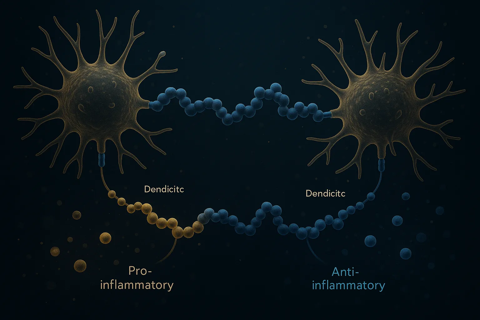
| Thymosin Alpha-1 | Immune system balancing (TLR/dendritic cell) | Immune dysregulation | Phase 3 + decades clinical use | Injectable |
| GHK-Cu | Gene modulation (4,000+ genes) | Tissue remodeling, chronic inflammation | Preclinical + human (topical) | Injectable/Topical |
1. KPV -- Direct NF-kB Pathway Inhibitor
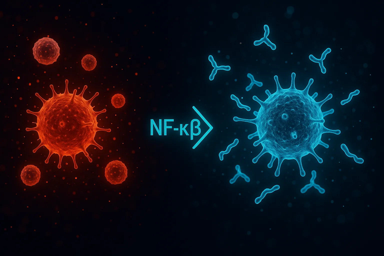
KPV (Lys-Pro-Val) is a C-terminal tripeptide derived from alpha-melanocyte-stimulating hormone that functions as one of the most potent direct inhibitors of NF-kB signaling available in peptide form. NF-kB is the master transcription factor controlling inflammatory gene expression -- it drives production of TNF-alpha, IL-1beta, IL-6, COX-2, and virtually every other major inflammatory mediator. Blocking NF-kB at the source is mechanistically upstream of what NSAIDs and most conventional anti-inflammatories achieve.
The mechanism is elegant: KPV enters cells and directly inhibits p65RelA nuclear translocation by stabilizing IkB-alpha (the endogenous NF-kB inhibitor). This blocks the inflammatory transcription factor from reaching the nucleus and activating its target genes. Competition assays revealed that KPV interacts with the importin-alpha3 binding site on p65RelA, physically preventing nuclear import [1]. This effect occurs at nanomolar concentrations -- orders of magnitude lower than most pharmaceutical anti-inflammatory agents.
Unlike the core alpha-MSH peptide, KPV does not appear to work through melanocortin receptors [2]. This is important because it means the anti-inflammatory effect is independent of the hormonal signaling that full-length MSH engages. KPV achieves targeted NF-kB inhibition without the melanocortin receptor-mediated effects on pigmentation, appetite, or sexual function.
The strongest preclinical evidence comes from intestinal inflammation models. KPV reduced colitis severity through PepT1-mediated uptake in intestinal epithelial cells, with dose-dependent inhibition of inflammatory cytokine production and preserved epithelial barrier function. Oral nanoparticle delivery of KPV efficiently alleviated ulcerative colitis in mouse models, suggesting that oral bioavailability -- often problematic for peptides -- is achievable for this small tripeptide.
For systemic inflammatory conditions, KPV's small size (only 3 amino acids) gives it favorable pharmacokinetic properties. It crosses cell membranes readily, has reasonable oral bioavailability, and distributes systemically after both oral and injectable administration.
The primary limitation is the absence of human clinical trials for any inflammatory condition. All current evidence is preclinical, though the consistency across multiple inflammation models (gut, skin, lung, systemic) and the well-characterized mechanism of action provide a strong biological rationale.
2. BPC-157 -- Tissue-Level Anti-Inflammatory and Repair
BPC-157's anti-inflammatory effects are intertwined with its tissue repair mechanisms. While it is better known as a healing peptide (see our joint pain ranking), its anti-inflammatory actions deserve specific attention because they address local tissue inflammation at the injury site rather than systemic inflammatory pathways.
BPC-157 reduces inflammatory cytokine production in damaged tissue while simultaneously promoting angiogenesis and growth factor signaling that accelerates the transition from inflammatory phase to proliferative phase of healing. This dual action -- dampening inflammation while promoting repair -- is what makes BPC-157 fundamentally different from conventional anti-inflammatories that suppress inflammation without supporting the resolution process.
In adjuvant arthritis models, BPC-157 reduced both the inflammatory arthritic process and the gastrointestinal damage caused by NSAID treatment of that arthritis [3]. This finding is particularly relevant for anyone currently managing chronic inflammation with NSAIDs -- BPC-157 may address both the underlying inflammatory condition and the iatrogenic damage from conventional treatment.
The angiogenic mechanism is itself anti-inflammatory in chronic conditions. Poor blood supply to damaged tissue perpetuates inflammation because inflammatory mediators and cellular debris cannot be cleared efficiently. By restoring vascular supply through VEGFR2 upregulation, BPC-157 enables the natural resolution of inflammation that insufficient blood flow prevents.
BPC-157's recent safety pilot in humans (intravenous infusion) confirmed a favorable safety profile [4]. Combined with decades of preclinical evidence across hundreds of studies, it represents one of the most extensively studied research peptides for tissue-level inflammatory conditions.
For protocols and vendor comparisons, see the BPC-157 page.
Top BPC-157 Vendors
Ranked by price, COA availability, and reputation
3. LL-37 -- Cathelicidin Immunomodulator
LL-37 is the only human cathelicidin antimicrobial peptide and functions as a dual-purpose immunomodulator: it directly kills pathogens while simultaneously modulating the inflammatory response to prevent excessive tissue damage from the immune system's own activity. This dual action makes it uniquely suited for inflammation driven by chronic infection, biofilm-associated conditions, or immune system overactivation in response to microbial triggers.
The antimicrobial properties are broad-spectrum: LL-37 kills bacteria, fungi, and certain viruses, and disrupts established biofilms [5]. This matters for inflammation because many chronic inflammatory conditions -- chronic sinusitis, periodontal disease, Lyme disease, certain gut dysbioses -- are driven by persistent microbial presence that sustains the inflammatory response. Eliminating the infectious trigger is the most direct way to resolve infection-driven inflammation.
The immunomodulatory component is equally important. LL-37 attenuates LTA-induced phosphorylation of p38MAPK and Akt, reducing TNF-alpha and IL-6 production in macrophages. It promotes epithelial wound repair and angiogenesis while simultaneously dampening excessive inflammatory cytokine release. In periodontal ligament cells, LL-37 was confirmed to be both anti-inflammatory and pro-apoptotic, clearing damaged cells while reducing inflammatory signaling.
LL-37 also increases epithelial barrier integrity -- it enhances lung epithelial cell stiffness and decreases transepithelial permeability, preventing bacterial invasion [6]. This barrier-strengthening effect is relevant for gut inflammation, respiratory inflammation, and any condition where compromised epithelial barriers allow microbial translocation that perpetuates inflammatory signaling.
The main limitation for LL-37 is that it addresses infection-driven inflammation specifically. For sterile inflammatory conditions (autoimmune, metabolic, age-related), LL-37's antimicrobial component is less relevant, though its immunomodulatory effects may still provide benefit. Human clinical trials specific to chronic inflammatory conditions have not been completed.