articlesApril 4, 2026·11 min read

Best Peptides for Joint Pain: 5 Ranked

BPC-157 reduced chronic knee pain in 7 of 12 patients for 6+ months. 5 joint peptides ranked by mechanism and evidence.

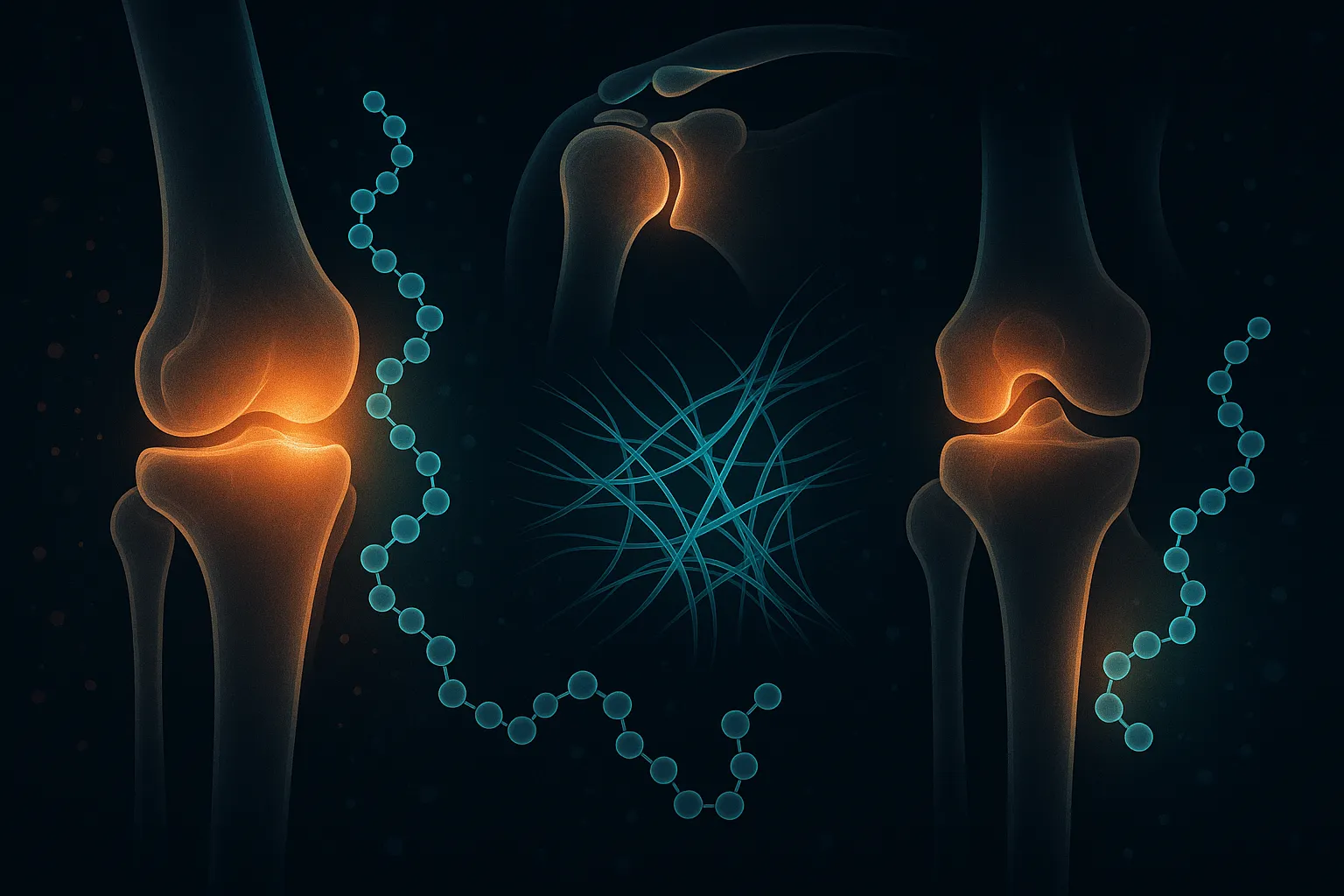
Best Peptides for Joint Pain: 5 Ranked (2026)

Best Peptides for Joint Pain

Joint pain affects over 90 million adults in the US alone, and conventional treatments -- NSAIDs, cortisone injections, physical therapy -- address symptoms without promoting actual tissue repair. Peptide therapy targets the underlying biology: stimulating angiogenesis at injury sites, recruiting stem cells, modulating inflammatory cascades, and supporting collagen synthesis in damaged connective tissue.

The challenge is navigating the evidence. Some peptides on this list have decades of preclinical research across hundreds of animal studies. Others have pilot human data that is encouraging but preliminary. And the gap between "reduces inflammation in a rat knee" and "repairs human osteoarthritis" is wider than most marketing suggests.

This article ranks 5 peptides for joint pain by their mechanism of action, strength of evidence, and practical application. Each section links to the full peptide page for detailed dosing protocols and vendor comparisons.

Quick Comparison Table

Peptide Mechanism Primary Target Evidence Level Route
BPC-157 Angiogenesis + growth factor signaling Tendons, ligaments, joints Extensive preclinical + pilot human Injectable (SC/local)
TB-500 Cell migration + anti-fibrosis Soft tissue, cartilage, muscle Preclinical + phase 2 (wound healing) Injectable (SC)
GHK-Cu Collagen synthesis + gene modulation Skin, connective tissue, joints Preclinical + human (topical) Injectable/Topical
KPV NF-kB inhibition + anti-inflammatory Systemic inflammation, gut Preclinical Oral/Injectable
Collagen peptides Structural substrate + chondrocyte stimulation Cartilage, synovial tissue Randomized human trials Oral

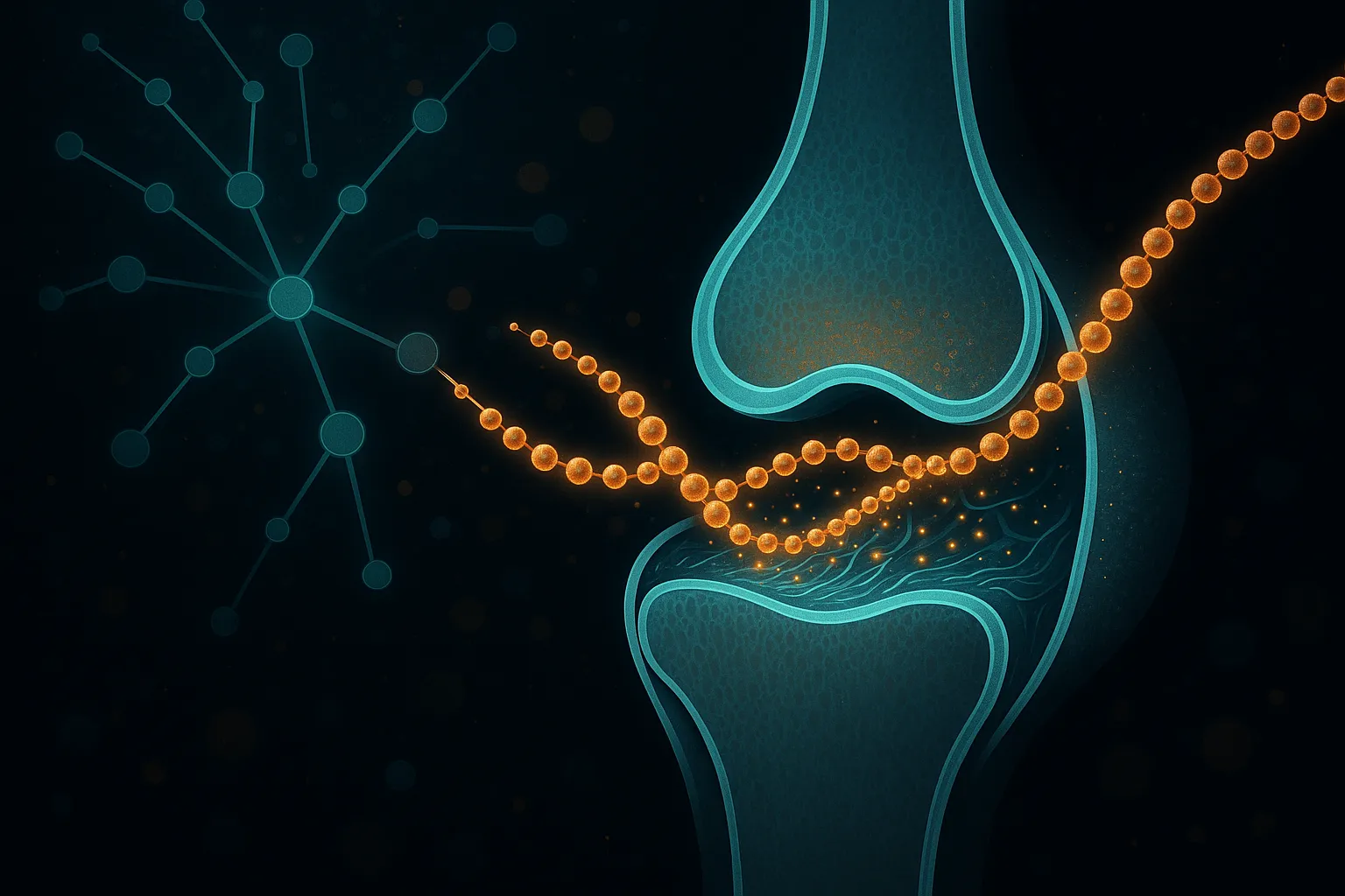
1. BPC-157 -- Gastric Pentadecapeptide

BPC-157 (Body Protection Compound-157) is a 15-amino-acid peptide derived from human gastric juice that has become the most widely researched peptide for musculoskeletal healing. Its mechanism centers on promoting angiogenesis through VEGFR2 upregulation, activating the FAK-paxillin pathway for cell migration, and stimulating growth hormone receptor expression in injured tissues.

The preclinical evidence is extensive. A 2021 study demonstrated that BPC-157 significantly accelerated tendon explant outgrowth and promoted tendon fibroblast survival and migration through FAK-paxillin pathway activation [1]. In Achilles tendon detachment models, BPC-157 improved functional recovery with substantially increased load-to-failure strength, better collagen fiber organization, and advanced vascular development at the repair site [2].

For joint-specific applications, BPC-157 has shown particular promise. A recent systematic review analyzed 36 studies (35 preclinical, 1 clinical) and found that BPC-157 improved functional, structural, and biomechanical outcomes across muscle, tendon, ligament, and bone injuries [3]. In the clinical study -- a pilot trial for chronic knee pain -- 7 of 12 patients reported pain relief lasting over 6 months after a single intra-articular BPC-157 injection. While this is a small, uncontrolled study, the durability of response from a single injection is notable.

BPC-157 also counteracts the joint-damaging effects of NSAIDs and corticosteroids. In adjuvant arthritis models, BPC-157 positively affected both NSAID-induced gastrointestinal lesions and the arthritic process itself, suggesting it may be particularly useful for individuals who need to transition away from conventional anti-inflammatory drugs [4].

The primary limitation is the absence of large-scale randomized controlled trials in humans. The preclinical evidence is among the strongest for any research peptide, but translating animal tendon and joint healing data to human osteoarthritis requires careful expectations.

Administration for joint applications typically involves subcutaneous injection near the affected joint or systemic subcutaneous injection. Some practitioners use direct intra-articular injection for severe cases. For full protocols, see the BPC-157 page.

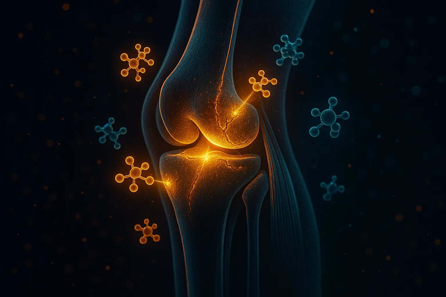
2. TB-500 (Thymosin Beta-4) -- Cell Migration and Anti-Fibrosis

TB-500 is a synthetic fragment of thymosin beta-4, a naturally occurring 43-amino-acid peptide found in virtually all human cells. Its primary mechanism for joint health involves promoting cell migration to injury sites, stimulating new blood vessel formation, and critically -- reducing fibrotic scar tissue formation that compromises joint function after injury.

The foundational research on thymosin beta-4 wound healing demonstrated that it accelerated wound closure by 42% at 4 days and 61% at 7 days compared to controls [5]. More relevant to joint applications, a phase 2 clinical trial in wound healing showed that thymosin beta-4 accelerated dermal healing by nearly a month in patients who responded, with mechanisms including stem cell mobilization, differentiation, and inflammation inhibition [6].

For connective tissue specifically, thymosin beta-4 enhanced medial collateral ligament healing in rats with improved structural and biomechanical properties. The anti-fibrotic effect is particularly relevant for joints: after injury, excessive scar tissue formation within the joint capsule restricts range of motion and predisposes to re-injury. TB-500's ability to organize connective tissue repair while preventing myofibroblast-driven scarring addresses this directly.

TB-500 also acts as a chemoattractant for myoblasts -- muscle progenitor cells -- following muscle injury. Since many joint pain presentations involve concurrent muscle weakness and atrophy around the affected joint (particularly knee and hip), this myoblast recruitment may support the broader functional recovery beyond just the joint structures.

The main limitation is that most human clinical data comes from wound healing rather than joint-specific trials. The translation from dermal wound repair to intra-articular tissue healing is reasonable given the shared cellular mechanisms (angiogenesis, cell migration, matrix remodeling) but is not directly proven.

TB-500 is administered via subcutaneous injection, typically at sites distant from the injury (systemic distribution). For dosing protocols and sourcing, see the TB-500 page.

3. GHK-Cu -- Copper Tripeptide

GHK-Cu (glycyl-L-histidyl-L-lysine copper complex) is a naturally occurring tripeptide present in human plasma, saliva, and urine that declines significantly with age. It functions as a master regulator of tissue remodeling, simultaneously stimulating collagen synthesis, glycosaminoglycan production, and controlled extracellular matrix turnover through metalloproteinase regulation.

The collagen synthesis evidence is robust. GHK-Cu stimulates both type I and type III collagen mRNA expression in fibroblasts at remarkably low concentrations (10^-12 to 10^-11 M), with collagen synthesis stimulation roughly double that of non-collagen proteins [7]. In wound chamber models, GHK-Cu produced concentration-dependent increases in total protein, collagen, and glycosaminoglycans -- the building blocks of cartilage and synovial tissue.

Beyond direct structural effects, GHK-Cu influences over 4,000 genes involved in tissue repair and inflammation. Gene expression studies show it suppresses genes associated with inflammation and tissue destruction while upregulating genes for antioxidant defense, DNA repair, and stem cell function [8]. This broad gene-modulatory effect distinguishes GHK-Cu from peptides that work through single receptor pathways.

For joint applications specifically, GHK-Cu's stimulation of decorin (a small proteoglycan critical for collagen fibril organization) and glycosaminoglycans (the main component of synovial fluid and cartilage matrix) makes it mechanistically relevant. The anti-inflammatory actions -- suppression of TNF-alpha, reduction of free radical damage, and inhibition of thromboxane formation -- address the inflammatory component of joint degeneration.

GHK-Cu is available in both injectable and topical formulations. For joint applications, subcutaneous injection near the affected area is most common. Topical application may support surrounding soft tissue but is unlikely to penetrate to intra-articular structures. For protocols and vendor comparisons, see the GHK-Cu page.

4. KPV -- Melanocortin Anti-Inflammatory Tripeptide

KPV (Lys-Pro-Val) is a C-terminal tripeptide fragment of alpha-melanocyte-stimulating hormone (alpha-MSH) that exerts potent anti-inflammatory effects through direct inhibition of the NF-kB signaling cascade -- the master regulator of inflammatory gene expression. Unlike full-length alpha-MSH, KPV does not appear to work through melanocortin receptors, instead entering cells and directly blocking p65RelA nuclear translocation.

Research demonstrates that nanomolar concentrations of KPV inhibit NF-kB activation and MAP kinase inflammatory signaling, reducing pro-inflammatory cytokine secretion including TNF-alpha and IL-6. The mechanism involves KPV entering the cell nucleus and stabilizing IkB-alpha (the NF-kB inhibitor), which prevents the inflammatory transcription factor from activating its target genes.

For joint applications, KPV's value lies in its broad anti-inflammatory action rather than direct tissue repair. Chronic joint pain is driven by sustained low-grade inflammation within the synovial membrane -- elevated TNF-alpha, IL-1beta, and IL-6 perpetuate cartilage degradation, synovial thickening, and pain sensitization. By targeting NF-kB directly, KPV addresses the upstream driver of this inflammatory cascade.

KPV has been most extensively studied in intestinal inflammation models, where it reduced colitis severity through PepT1-mediated uptake in epithelial cells. The translation to joint inflammation is mechanistically sound (NF-kB drives both gut and joint inflammation) but has not been directly tested in joint-specific models.

KPV is available in both oral and injectable formulations. Its small size (tripeptide) gives it favorable oral bioavailability compared to larger peptides. For systemic anti-inflammatory effects relevant to joint pain, either route may be effective.

5. Collagen Peptides -- Structural Support and Chondrocyte Stimulation

Collagen peptides (hydrolyzed collagen) represent the most conventional and best-studied oral option for joint support, with multiple randomized controlled trials in human joint pain populations. Unlike the research peptides above, collagen peptides have completed human efficacy trials specifically for joint outcomes.

A 24-week randomized trial of 147 athletes with activity-related joint pain found that 10 g daily of collagen hydrolysate significantly reduced pain assessed by a physician compared to placebo, with the most pronounced effects in knee and ankle joints. Multiple meta-analyses confirm modest but statistically significant improvements in joint pain, stiffness, and physical function scores in osteoarthritis populations with collagen supplementation.

The mechanism involves two components. First, collagen peptides provide the structural amino acids (glycine, proline, hydroxyproline) needed for cartilage matrix synthesis. Second, specific collagen-derived dipeptides (particularly Pro-Hyp) act as signaling molecules that stimulate chondrocyte biosynthesis -- increasing collagen and proteoglycan production by the cells responsible for maintaining cartilage integrity.

The effect sizes are modest compared to what the peptide therapeutics above promise in preclinical models. But collagen peptides have something the others largely lack: completed, peer-reviewed human trials specifically measuring joint pain outcomes in relevant populations. The safety profile is excellent with no significant adverse effects reported in any trial.

Collagen peptides are taken orally at 10-15 g per day, typically as a powder dissolved in liquid. They are widely available, inexpensive, and require no injection. For individuals starting joint support, collagen peptides represent the lowest-risk entry point.

Stacking Strategies for Joint Pain

The most common and mechanistically sound joint pain stack combines BPC-157 and TB-500. BPC-157 drives angiogenesis and growth factor signaling at the injury site while TB-500 promotes cell migration, stem cell recruitment, and anti-fibrotic tissue remodeling. These complementary pathways address different phases of the healing cascade simultaneously.

  • BPC-157 + TB-500: The foundational joint healing stack. Run both for 8-12 weeks during active injury recovery. BPC-157 typically at 250-500 mcg/day subcutaneous, TB-500 at loading dose then maintenance.
  • BPC-157 + GHK-Cu: Add GHK-Cu when collagen remodeling is a priority -- chronic tendinopathy, post-surgical recovery, or degenerative changes where the extracellular matrix needs rebuilding. GHK-Cu's collagen synthesis stimulation complements BPC-157's vascular and growth factor effects.
  • Any injectable protocol + collagen peptides: Oral collagen peptides provide the raw structural substrates (glycine, proline, hydroxyproline) that the body needs to build new connective tissue. Adding 10-15 g daily of collagen hydrolysate during any peptide-based joint protocol ensures the building blocks are available.
  • KPV + BPC-157: For joints where chronic systemic inflammation is the primary driver (rheumatoid arthritis, psoriatic arthritis, systemic inflammatory conditions), adding KPV to target NF-kB while BPC-157 handles local tissue repair addresses both systemic and local components.

Joint Healing Mechanisms

How to Choose: Decision Framework

Acute injury recovery (sports injury, post-surgical): BPC-157 + TB-500 is the standard combination. The goal is accelerating the natural healing cascade -- angiogenesis, cell migration, matrix deposition -- during the critical window when tissue remodeling is most active.

Chronic degenerative joint pain (osteoarthritis, chronic tendinopathy): BPC-157 as the primary agent, with GHK-Cu added for collagen remodeling support. Chronic conditions require longer protocols (12-16 weeks) and expectations should focus on pain reduction and functional improvement rather than structural reversal.

Systemic inflammatory joint conditions: KPV for upstream NF-kB inhibition combined with BPC-157 for local tissue effects. Individuals with inflammatory arthritis or autoimmune-driven joint pain need the systemic anti-inflammatory component that BPC-157 alone may not adequately provide.

Low-risk starting point: Oral collagen peptides at 10-15 g/day. Proven in human trials, no injections, excellent safety profile. Combine with any injectable protocol as a substrate foundation.

Budget-conscious: BPC-157 alone is effective for many joint conditions and costs $40-80/month from research vendors. TB-500 adds $40-60/month. Collagen peptides run $15-30/month.

Recovery Timeline Expectations

Joint healing is slow tissue. Unlike muscle, which has robust blood supply and rapid turnover, cartilage, tendons, and ligaments have limited vascularity and slow cellular metabolism. Even with peptide support:

  • Weeks 1-2: Reduced acute inflammation and early pain relief
  • Weeks 2-4: Improved joint stiffness and range of motion
  • Weeks 4-8: Functional improvements in daily activities
  • Weeks 8-12: Connective tissue remodeling and structural adaptation
  • Weeks 12-16+: Maximum benefit for chronic conditions

These timelines apply to most connective tissue injuries. Acute injuries with good blood supply (muscle tears near joints, acute tendinitis) respond faster. Chronic degenerative conditions (advanced osteoarthritis, calcified tendons) respond slower and may require multiple protocol cycles.

Joint Recovery Timeline

Monitoring and Assessment

Joint peptide protocols benefit from structured progress tracking:

  • Pain scores: Track daily pain on a 0-10 scale at consistent times (morning stiffness, end-of-day activity pain). Weekly averages are more useful than single data points.
  • Range of motion: Measure and record joint-specific ROM at baseline and every 2 weeks. Goniometer measurements for knees and shoulders; finger-to-floor distance for spinal mobility.
  • Functional tests: Timed sit-to-stand, stair descent pain rating, grip strength (for hand/wrist joints). These objective measures complement subjective pain scores.
  • Imaging (if available): Ultrasound can track tendon thickness, synovial fluid volume, and neovascularization at 8-12 week intervals. MRI at baseline and 16 weeks for significant structural injuries.

References

  1. Chang CH, et al. The promoting effect of pentadecapeptide BPC 157 on tendon healing involves tendon outgrowth, cell survival, and cell migration. J Appl Physiol. 2011;110(3):774-780. PMID: 21030672
  2. Cerovecki T, et al. Pentadecapeptide BPC 157 (PL 14736) improves ligament healing in the rat. J Orthop Res. 2010;28(9):1155-1161. PMID: 20225319
  3. Kang EA, et al. Emerging use of BPC-157 in orthopaedic sports medicine: a systematic review. Am J Sports Med. 2025. PMID: 40756949
  4. Sikiric P, et al. Pentadecapeptide BPC 157 positively affects both non-steroidal anti-inflammatory agent-induced gastrointestinal lesions and adjuvant arthritis in rats. J Physiol Paris. 1997;91(3-5):113-122. PMID: 9403784
  5. Malinda KM, et al. Thymosin beta4 accelerates wound healing. J Invest Dermatol. 1999;113(3):364-368. PMID: 10469335
  6. Goldstein AL, et al. The regenerative peptide thymosin beta4 accelerates the rate of dermal healing in preclinical animal models and in patients. Ann N Y Acad Sci. 2012;1270:37-44. PMID: 23050815
  7. Maquart FX, et al. Stimulation of collagen synthesis in fibroblast cultures by the tripeptide-copper complex glycyl-L-histidyl-L-lysine-Cu2+. FEBS Lett. 1988;238(2):343-346. PMID: 3169264
  8. Pickart L, et al. Regenerative and protective actions of the GHK-Cu peptide in the light of the new gene data. Int J Mol Sci. 2018;19(7):1987. PMID: 29986520